|
The shaft from which this steel specimen was made fractured
in service.
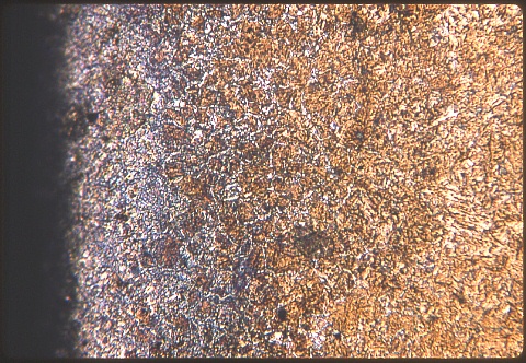
I made first four photomicrographs at 200X each. They progress
from the very edge (on the left side of the first image) inwards to the
center of the shaft.
The microconsituents are rather finely dispersed and are difficult to
resolve, so I'll help you out a little:
* At the very edge there exist ferrite and fine pearlite, which grade
to 100% martensite towards the interior.
|
|
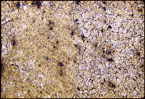
* In the second image, farther from the the edge, the
gradation is from all martensite at the left to martensite plus fine
pearlite.
|
|
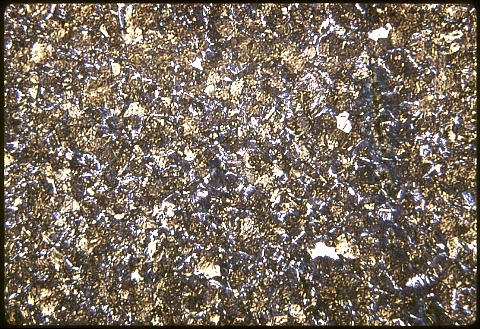
* In the third image, halfway along the radius of the shaft,
there is massive cementite, pearlite, and martensite.
|
|
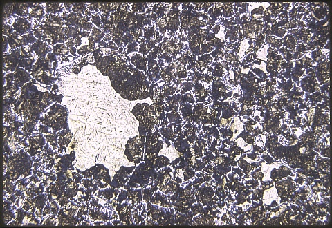
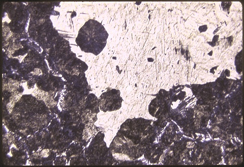
* The fourth image shows one large area and
several smaller patches of martensite in a matrix of cementite plus
pearlite.
|
|
* The last image shows another patch of
martensite with somewhat better resolution at 500X magnification.
Write
out your answer; complete all or part of the set; and then apply for a
Username
& Password so you can activate the link at the bottom of this page.
|